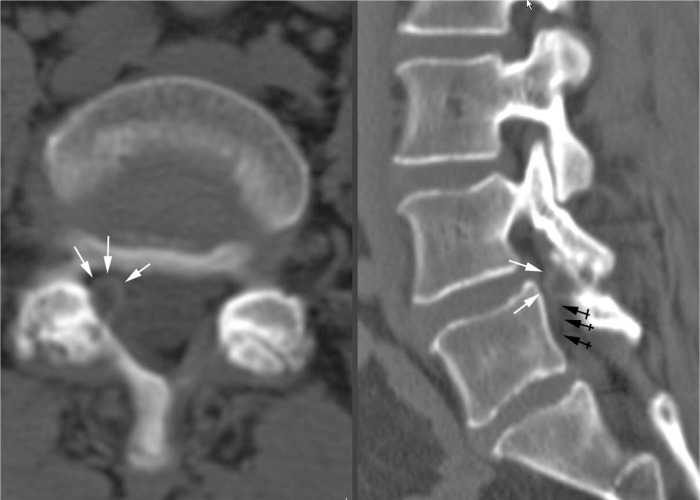
Figure 2.
Axial and reformatted right parasagittal CT views in bone windows show a juxtaarticular cyst with a thin rim of attenuated density (white arrows), occluding the right L5 lateral recess and compressing the right L5 nerve root (black arrows) anterior to a hypertrophied and narrowed right L4/5 facet joint with subchondral cystic changes.

