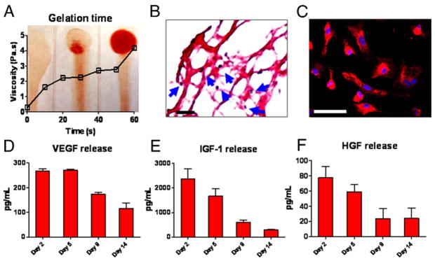
Figure 1. Characterization of Platelet Gel In Vitro.

(A) Mixture of platelet-containing plasma and Dulbecco’s Modified Eagle’s Medium was immediately dispensed onto coverslips, and gel formation was monitored over time. At 0 s, tilting the coverslip resulted in a liquid smear, indicating no gel formation. Gel formation started at 30 s, and at 60 s, only a little smear was seen when tilting the coverslip, indicating gelation. Viscosity was measured by a rheometer and plotted against time. The increase of viscosity over time was consistent with the progress of gelation. (B) Hematoxylin-eosin staining revealed a fibrous structure of the platelet gel and the presence of platelets (blue arrows). Bar = 100 um. (C) Neonatal rat cardiac myocytes (NRCM) cultured in the platelet gel for 14 days and stained with CM-DiI. Bar = 50 um. (D to F) Concentrations of vascular endothelial growth factor (VEGF), insulin-like growth factor (IGF)-1, and hepatocyte growth factor (HGF) from platelet gel-conditioned media at various time points (n = 3 per time point) measured by enzyme-linked immunosorbent assay.
