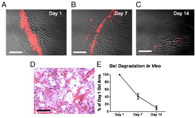
Figure 2. Degradation of Injected Platelet Gel in Infarcted Rat Hearts.

Platelet gel labeled with Texas Red-X succinimidyl ester was intramyocardially injected into Wistar-Kyoto rat hearts with myocardial infarction (MI), and heart sections were studied histologically at various time points. (A to C) The areas of platelet gel could be readily identified with the detection of Texas Red fluorescence. Bars = 500 um. (D) Hematoxylin-eosin staining revealed penetration of endogenous cells into the gel. Bar = 50 um. (E) To calculate the percentage of degradation over time, the gel area at days 7 and 14 was measured and then normalized to the day 1 gel area.
