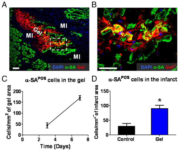
Figure 3. Cardiomyocytes Populate Injected Platelet Gel In Vivo.
Animals were sacrificed at days 3 and 7, and heart cryosections were stained with 4′,6-diamidino-2-phenylindole (DAPI) for nuclei and alpha-sarcomeric actin (α-SA) for cardiomyocytes. (A) Representative confocal microscopic images. The infarct area could be identified by the paucity of cardiomyocytes and endothelial cells but with infiltration by nuclei belonging to inflammatory cells (marked as “MI”). Platelet gel exhibited Texas Red fluorescence (marked as “Gel”). (B) High-magnification image of the area in the white box in A, showing the penetration of cardiomyocytes into the platelet gel. (C) Quantitation of myocytes residing in the platelet gel at day 3 and day 7, respectively. (D) Quantitation of cardiomyocytes in the infarct area from gel- or control-treated hearts on day 7 after treatment. Bars = 100 um. *p < 0.0001 when compared to control. POS = positive.

