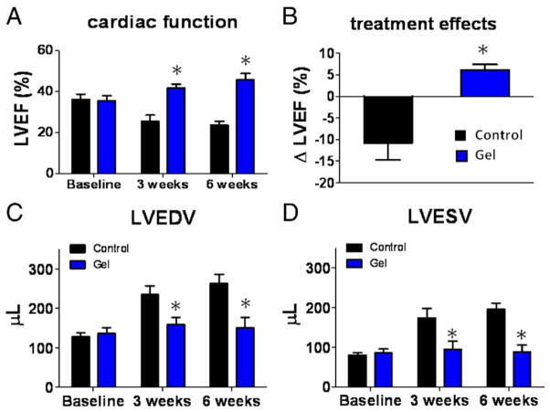
Figure 6. Cardiac Function and Chamber Dimensions.
(A) Left ventricular ejection fraction (LVEF) measured by echocardiography at baseline (post-myocardial infarction), 3 weeks and 6 weeks afterward in control group (black bars) or gel-treatment group (blue bars) (n = 8 animals per group). Baseline LVEFs were indistinguishable in the 2 groups. (B) Changes of LVEF from baseline to 3 weeks in each group. (C) Left ventricular end-diastolic volumes (LVEDV) measured by echocardiography. (D) Left ventricular end-systolic volumes (LVESV) measured by echocardiography. *Indicates p < 0.05 when compared to control.

