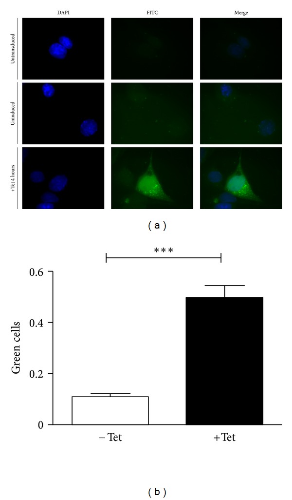
Figure 2.

Inducible AIFsh expression in cardiac cells. HL-1 cardiac cells were transduced with the pNRTA vector and assessed for transgene expression 48 hours later. Transgene expression was evaluated before or after induction by exposing cells to 1 μg/mL Tet for 4 hours. (a) Cells were then processed for immunofluorescence with an anti-AIF antibody and an FITC-conjugated secondary antibody. The panel shows sample images of HL-1 cells treated as follows: untransduced (upper), transduced and uninduced (middle), and transduced and induced (lower). DAPI staining, green fluorescence, and a merging of the two channels are shown. (b) Quantitation of immunofluorescent slides of transduced HL-1 cells either uninduced (−Tet) or induced (+Tet). The amount of fluorescent cells is expressed as a fraction of green fluorescent cells over nonfluorescent cells in the field counted.
