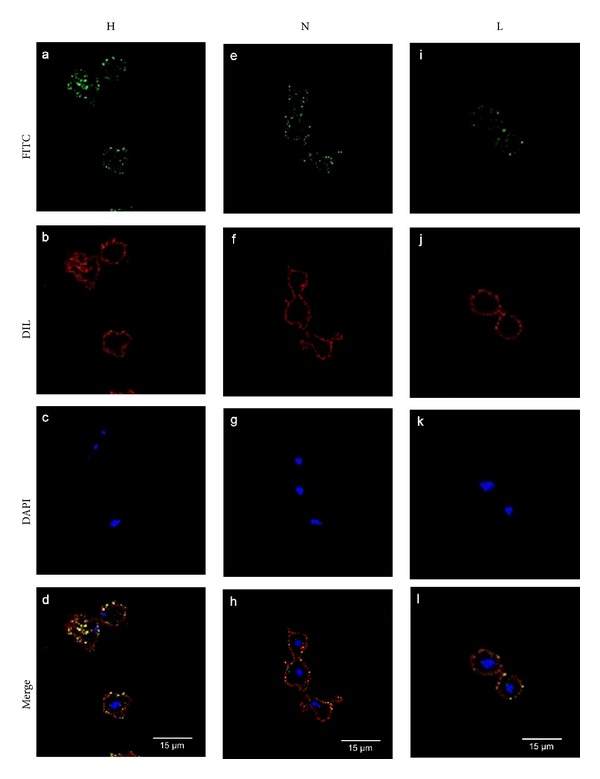
Figure 3.

The TvLEGU-1 surface localization on T. vaginalis is affected by iron. Parasites grown in iron-rich (H; a, b, c, and d), normal (N; e, f, g, and h), and iron-depleted (L; i, j, k, and l) conditions, fixed and nonpermeabilized were incubated with the anti-TvLEGU-1r antibody (1 : 100 dilution). Anti-rabbit IgG-FITC (in green) was used as a secondary antibody (1 : 100 dilution) (a, e, and i). Parasite membranes were labeled with DIL (in red; b, f, and j). Nuclei were labeled with DAPI (in blue; c, g, and k). Merge (d, h, and l) in yellow indicates colocalization. Bars: 15 μm (d, h, and l).
