Figure 9.

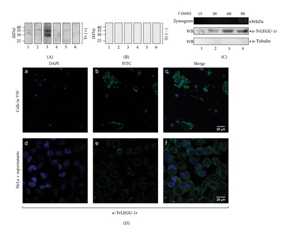
Presence of TvLEGU-1 in vaginal secretions and in in vitro secretion assays. (A) WB assays of TCA-precipitated proteins present in VWs from T. vaginalis positive culture patients [Tv (+)] (lanes 1–6) incubated with the anti-TvLEGU-1r antibody. (B) WB assays of TCA-precipitated proteins from people with other vaginitis [Tv (−)] used as negative controls (lanes 1–6) incubated with the anti-TvLEGU-1r antibody. (C) Zymogram and WB assays of the proteins present in the in vitro secretion products obtained from metabolically active parasites (1 × 10⁶ cells/mL) that were incubated in PBS-0.5% maltose at 37°C for 15, 30, 60, and 90 min (lanes 1–4, resp.). NC membranes containing the TCA-precipitated in vitro secretion products incubated with the anti-TvLEGU-1r antibody (1 : 10,000) or the anti-α-tubulin antibody (1 : 1000) used as a negative control. (D) Confocal microscopy images of fixed cells obtained from vaginal washes (VWs) and from live HeLa cell. (a, b, and c) Cells from VWs of patients with trichomoniasis confirmed by in vitro culture [Tv (+)] directly incubated with the anti-TvLEGU-1r antibody. (d, e, and f) Live HeLa cells incubated with supernatants from the in vitro secretion assays in which TvLEGU-1 is present (C) and with the anti-TvLEGU-1r antibody. Conjugated anti-rabbit IgG-FITC was used as a secondary antibody (1 : 100 dilution) (b and e). Nuclei labeled with DAPI (a and d); merge, and bars: 20 μm (c and f).
