FIG. 1.
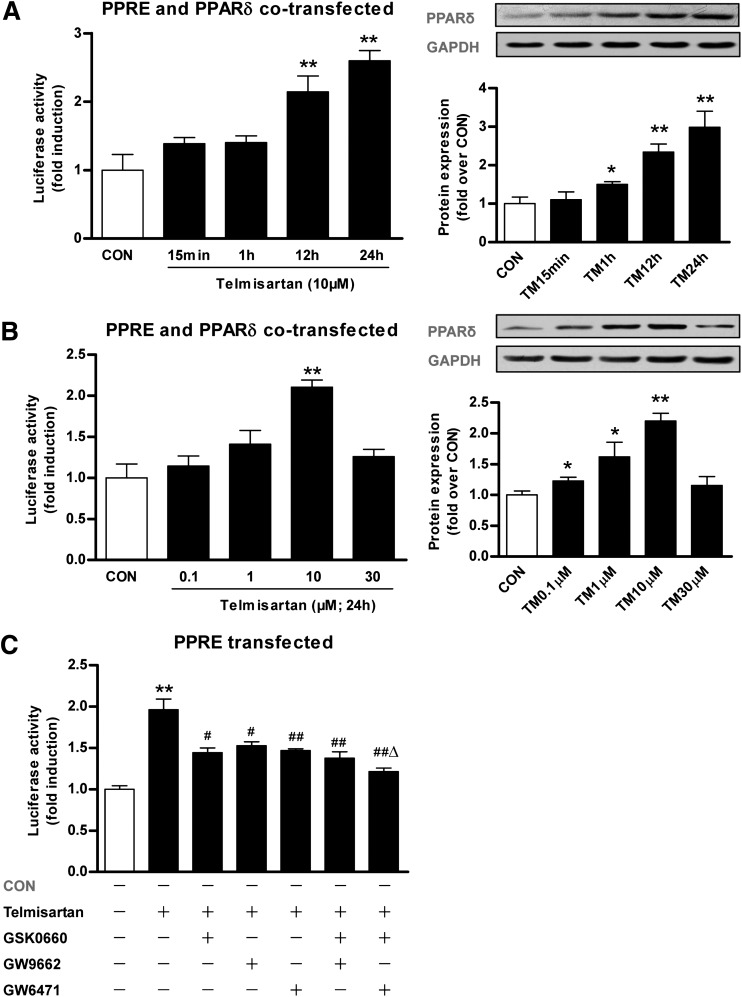
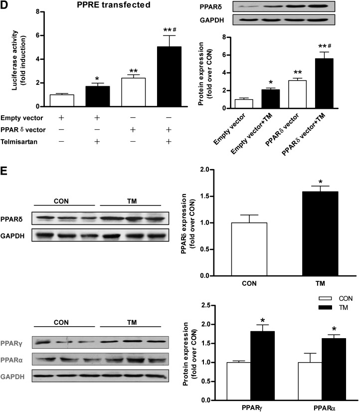
Effect of TM on PPARδ expression and activity in C2C12 myotubes. A: PPRE activity in C2C12 myotubes transfected with pTK-PPREx3-luc was evaluated by luciferase assay. PPARδ expression in C2C12 myotubes transfected with pTK-PPREx3-luc was detected by Western blotting. C2C12 myotubes with cotransfection the plasmid pAdTrack-CMV-PPAR-δ, containing the full-length coding region of rat PPARδ (PPARδ vector), were treated with DMSO (control [CON]) or TM (10 μmol/L) for 15 min to 24 h before the luciferase assay or Western blotting. *P < 0.05 and **P < 0.01 vs. CON. B: PPRE activity in C2C12 myotubes cotransfected with pTK-PPREx3-luc and PPARδ vector was evaluated by luciferase assay. PPARδ expression in C2C12 myotubes transfected with pTK-PPREx3-luc was detected by Western blotting. Cells were treated with DMSO (CON) or indicated concentrations of TM (0.1–30 μmol/L) for 24 h. *P < 0.05 and **P < 0.01 vs. CON. C: Cells were treated with DMSO (CON) or 10 μmol/L TM for 24 h. Cells were treated with 10 μmol/L TM in the presence or absence of PPARδ inhibitor GSK0660 (GSK; 10 μmol/L), PPARγ inhibitor GW9662 (GW9662; 10 μmol/L), or PPARα inhibitor GW6471 (GW6471; 10 μmol/L) for 24 h before the luciferase assay. **P < 0.01 vs. CON; #P < 0.05 and ##P < 0.01 vs. TM 10 μmol/L; ΔP < 0.05 vs. TM 10 μmol/L plus GSK 10 μmol/L. D: 10 μmol/L TM treatment for 24 h increased the luciferase activity both in an empty pAdTrack expression vector as the control and the cotransfection with the PPARδ expression vector in transfected C2C12 myotubes. *P < 0.05 and **P < 0.01 versus CON; #P < 0.05 vs. PPARδ vector plus TM 10 μmol/L. E: PPARδ, PPARγ, and PPARα expression in C2C12 myotubes was detected by Western blotting after treatment with DMSO (CON) or TM (10 μmol/L) for 24 h. *P < 0.05 vs. CON. Data are mean ± SEM from 3–6 experiments.


