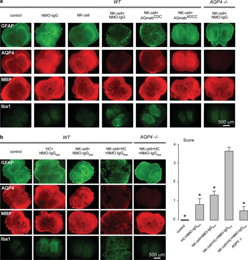
Fig. 6.
NK-cells exacerbate NMO lesions produced by NMO-IgG and complement in an ex vivo spinal cord slice culture model. a Immunofluorescence of GFAP, AQP4, MBP and Iba1 in spinal cord slice cultures from wild type (WT) and AQP4-/- mice incubated with NMO-IgG, AQmabCDC or AQmabADCC (each 10 μg/mL) and/or NK-cells (106/well). Control indicates no NMO-IgG or NK-cells. b Same staining as in (a) of spinal cord slice cultures incubated with NK-cells (106/well) and/or human complement (HC, 5 %) and/or submaximal NMO-IgG (NMO-IgGlow, 3 μg/mL) (left). Scoring of NMO lesions (mean ± SE, 6–8 slices per condition, *P < 0.001 compared with NK-cell + HC + NMO-IgGlow, WT) (right)

