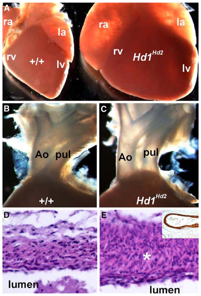
Fig. 2.

Cardiac defects in the high-percentage Hand1Hand2 chimeric newborns. a–c Wild-type (+/+) and stillborn high-percentage Hd1Hd2 chimeric age-matched littermate hearts. Note the dilation of both atria and ventricles and the rounded apex at the base of the heart compared with controls. Closer examination shows that high-percentage chimeras exhibit double-outlet right ventricle (DORV) as both the aorta (Ao) and pulmonary trunk (pul) exit the right ventricle (c) compared with wild-type littermates (b). d, e Hematoxylin and eosin (H&E) sections through the wild-type (d) and Hd1Hd2 chimeric aortic arch show hyperplasia of the smooth muscle layer (asterisk) around the outflow tract (OFT) vessels. Note however, that there is appropriate α-smooth muscle actin (αSMA) staining of the Hd1Hd2 chimeric vasculature (shown in the inset in e). ra right atria, rv right ventricle, la left atria, lv left ventricle
