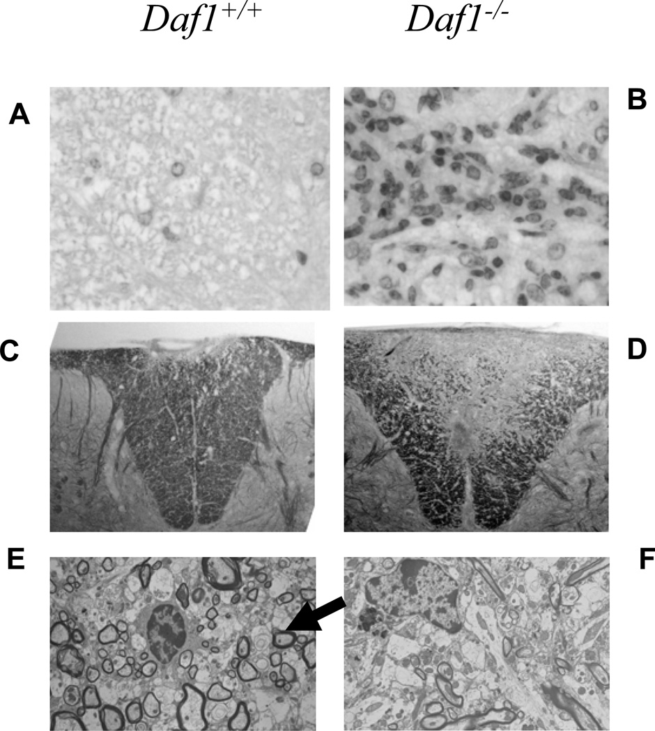
Figure 2. Neuropathology of spinal cords in MOG35–55 immunized Daf1−/− and Daf1+/+ mice.
A and B) H&E staining of spinal dorsal column (magnification X100). C and D) Luxol fast Blue stains of dorsal column sections (magnification X100). E and F) Electron microscopy of lesions in dorsal columns at day 84 post immunization. E) The arrow shows thinly remyelinated axons in Daf1+/+ mice.

