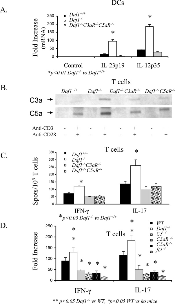
Figure 6. Augmented T cell responses by Daf deficient APCs and T cells depend on C5aR and C3aR.
A) IL-23p19 and IL-12p35 mRNA expression in sorted Daf1+/+, Daf1−/−, and Daf1−/−C3aR−/−C5aR−/− DCs after incubation with ova323–339 and OT-II cells. B). Western Blots of C3a and C5a in supernatants of Daf1+/+, Daf−/−, Daf1−/−C3aR−/− and Daf1−/−C5aR−/− T cells following anti-CD3 and anti-CD28 antibody stimulation. C) IFN-γ and IL-17 ELISPOT assays of Daf1+/+, Daf1−/−, Daf1−/− C5aR−/−, and Daf1−/−C3aR−/− T cells following anti-CD3 and anti-CD28 antibody stimulation. D) IFN-γ and IL-17 protein levels in supernatants of anti-CD3 and anti-CD28 antibody stimulated T cells from WT and relevant knockout mice. T cell were isolated by CD3+ T cell enrichment columns (R&D Systems).

