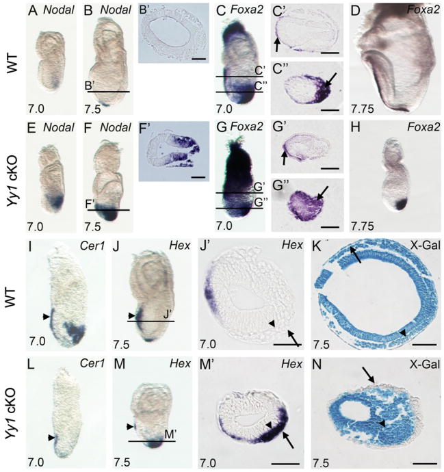
Fig. 3.
Defective gastrulation in Yy1 cKO mutants. WISH of Nodal (A–B and E–F) showing failure of spatiotemporal repression in Yy1 mutants. B′ and F′ are transverse sections corresponding to the lines in B and F. WISH of Foxa2 (C–D and G–H). C, C″, G′ and G″ are transverse sections corresponding to the lines in C and G. WISH of Cer1 (I and L); Hex (J and M) marking properly migrating AVE cells in WT (arrowhead in J) and mutant embryos (arrowhead in M). In WT embryos, Hex positive DE cells have intercalated into VE and have migrated anteriorly. Mutant Hex positive cells (arrowhead in M′) fail to intercalate into VE (arrow in M′). J′ and M′ are transverse sections corresponding to the lines in J and M. Arrows in J′ and M′ indicate VE. Transverse sections of X-gal stained R26R WT (K) and mutant (N) embryos. Arrowhead in K and N indicate epiblast derivatives. Arrows in K and N indicate cells in the outer layer of the conceptus, showing β-gal positive epiblast derived DE in WT embryos and β-gal negative extraembryonic derived VE in mutants. Scale bars represent 75 mm.

