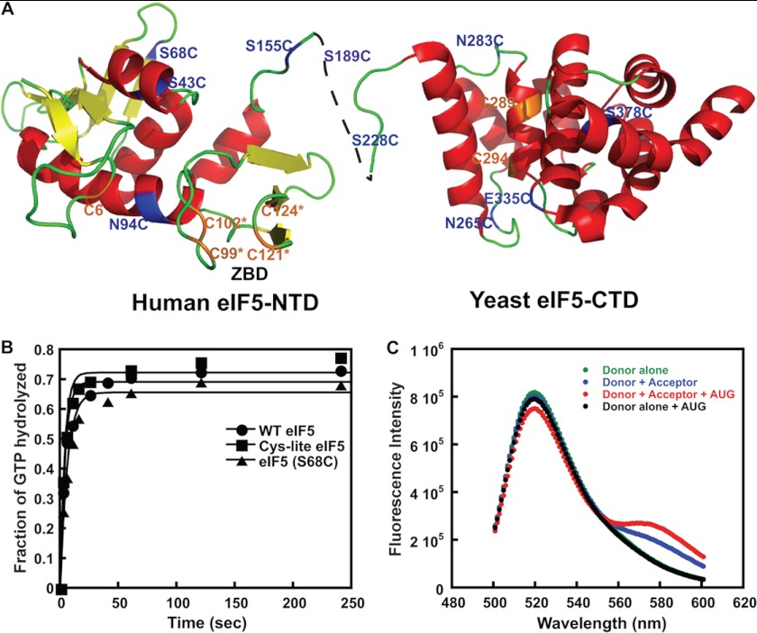
FIGURE 1.
FRET between fluorophores in the NTD of eIF5 and CTD of eIF1A in the PIC upon AUG recognition. A, ribbon representation of the human eIF5 NTD and yeast eIF5 CTD showing the single cysteine mutants generated in the Cys-lite background. Positions of native cysteines (C6, C289, and C294) that are not involved in the ZBD are shown in orange. Cysteines introduced at non-conserved surface residues are shown in blue. The cysteines involved in the ZBD (C99*, C102*, C121*, and C124*) are shown in orange. B, kinetics of GTP hydrolysis by eIF2, performed as described under “Experimental Procedures,” in the presence of native eIF5 (closed circles), Cys-lite eIF5 (closed squares), and Cys-lite eIF5(S68C) (closed triangles). Points are averages from two independent experiments. C, steady-state fluorescence measurements demonstrating FRET in the PIC upon the addition of mRNA(AUG) between the Cys-lite eIF5(S68C) derivative labeled with fluorescein and eIF1A labeled C-terminally with TAMRA. The following complexes were assembled, and their fluorescence was measured as a function of emission wavelength (excitation wavelength = 490 nm): Cys-lite eIF5(S68C)-Fl·eIF1A·eIF1·40 S·TC (Donor alone; green); eIF5(S68C)-Fl·eIF1A-TAMRA·eIF1·40 S·TC (Donor + Acceptor; blue); eIF5(S68C)-Fl·eIF1A-TAMRA·eIF1·40 S·TC·mRNA(AUG) (Donor + Acceptor + AUG; red); eIF5(S68C)-Fl·eIF1A·eIF1·40 S·TC·mRNA(AUG) (Donor alone + AUG; black). The FRET change can be seen as both a decrease in fluorescein (donor) fluorescence at 520 nm and increase in TAMRA (acceptor) fluorescence at 580 nm upon the addition of mRNA(AUG) to the donor + acceptor complex (red versus blue curves). The emission seen at 580 nm in the Donor + Acceptor curve in the absence of mRNA (blue) is due to weak excitation of TAMRA by the incident light. No change in donor fluorescence is observed in the absence of acceptor upon the addition of mRNA(AUG) (green versus black curves), demonstrating that the decrease in emission is due to FRET rather than a change in intrinsic fluorescence of the fluorescein moiety.

