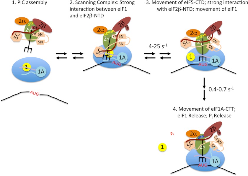
FIGURE 6.
Model for the events taking place within the PIC upon start codon recognition. Stage 1, TC·eIF5 complex binds to the 40S subunit. eIF1 occupies a site on the platform of the 40S adjacent to the P site, and the body of eIF1A binds in the A site, with its NTT (purple) and CTT (light blue) binding in the P site. Stage 2, the scanning PIC is in an open conformation with the tRNAi in the Pout state. eIF1 binding is stabilized by a strong interaction with the NTT of eIF2β. Stage 3, entry of the start codon into the P site allows formation of the codon-anticodon helix between the mRNA and tRNAi, which drives the tRNA into the Pin state. This displaces eIF1 to a second, weaker binding site on the 40S subunit, breaking its interaction with the eIF2β NTT, which in turn binds strongly to the eIF5 CTD. Movements of the tRNA and/or eIF5 CTD result in changes in the orientation of the eIF5 NTD. Stage 4, eIF1 dissociates from the complex, which, along with accommodation of tRNAi into the P site, causes the CTT of eIF1A to move and interact with the eIF5 NTD. This interaction triggers Pi release from eIF2, possibly by moving the unstructured NTT of eIF5 (not shown for clarity). The resulting complex is in a closed, scanning-arrested state.

