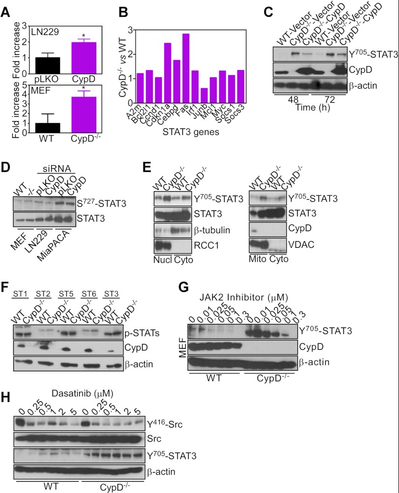
FIGURE 5.
Selective STAT3 activation induced by CypD loss. A, LN229 transfected with control (Ctrl), CypD-directed shRNA (top panel), or WT or CypD−/− MEFs (bottom panel) were transfected with a STAT3 luciferase reporter construct and analyzed for promoter activity in a luminometer after 24 h. Data are mean ± S.D. (n = 3). B, RNA extracted from WT or CypD−/− MEFs was amplified with primers specific for the indicated STAT3-regulated gene products by PCR. Data are expressed as fold differences in mRNA expression normalized to WT MEFs. C, WT or CypD−/− MEFs were transfected with the indicated plasmids and analyzed by Western blotting. D, the indicated cell types were analyzed by Western blotting. E, WT or CypD−/− MEFs were fractionated in the indicated subcellular compartments and analyzed by Western blotting. Nucl, nuclei; Cyto, cytosol; Mito, mitochondria. VDAC or RCC1 were used as mitochondrial or nuclear markers, respectively. F, WT or CypD−/− MEFs were analyzed for changes in expression of various STAT molecules by Western blotting. G, WT or CypD−/− MEFs were incubated with the indicated increasing concentrations of a JAK2 inhibitor and analyzed by Western blotting. H, WT or CypD−/− MEFs were treated with the indicated increasing concentrations of Dasatinib and analyzed by Western blotting. The phosphorylation sites in Src or STAT3 are indicated.

