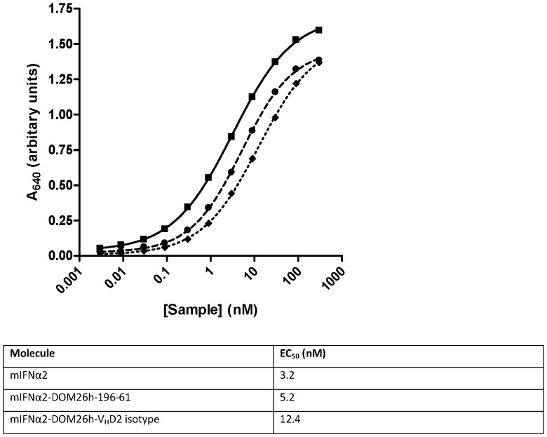
Figure 4. In vitro activity of mIFNα2 formatted as dAb fusions.
Activity of the mIFNα2-dAb fusion proteins was tested in the B16-Blue™ assay and compared to unfused mIFNα2 standard. Error bars are not visible as they are smaller than the data points, but represent standard error of the mean of 3 independent experiments. mIFNα2-DOM26h-196-61 (dashed line, closed circles) and mIFNα2-VHD2 isotype control (dotted line, closed diamonds) showed comparable activity to the H6-mIFNα2 standard (solid line, closed squares), with only minor increases in the EC50.

