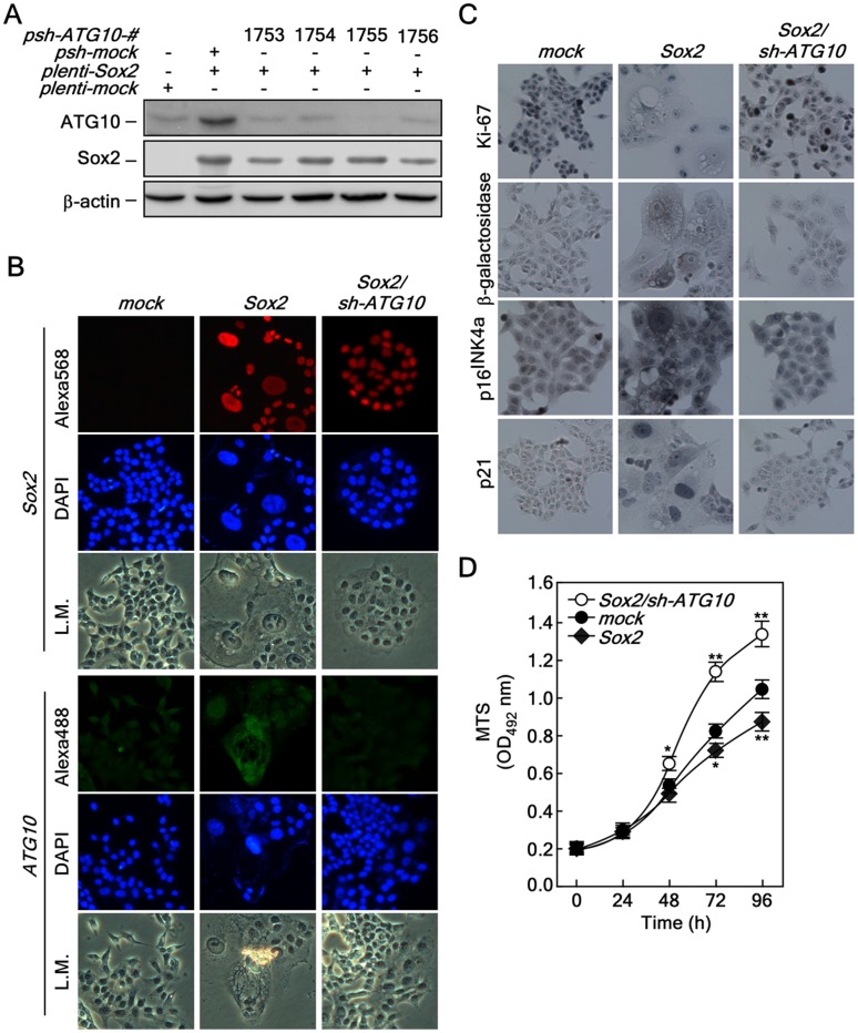
Figure 6. Knockdown of ATG10 restores Sox2-induced autophagy, cellular senescence and proliferation.
(A) Confirmation of the knockdown efficiency of ATG10. Knockdown of ATG10 using pLKO-shATG10. The pLKO-shATG10 knockdown lenti-viral particles were infected into HCT116 cells stably expressing Sox2 and cells cultured for 36 h. The proteins were extracted and knockdown efficiency was determined by Western blot using a specific ATG10 antibody. ß-Actin was used as an internal control to verify equal protein loading. (B) Morphological changes induced by ATG10 knockdown in HCT116 cells stably expressing Sox2. HCT116-mock, -Sox2 and -Sox2/sh-ATG10 cells were analyzed for morphological changes and protein levels of Sox2 (red) and ATG10 (green) were determined by an immunofluorescence assay using specific antibodies. The cells were observed under a fluorescence or light microscope (X200). L.M. indicates light microscopy. (C) Restoration of cell cycle, cell proliferation and senescence markers by knocking down ATG10 in HCT116 cells stably expressing Sox2. HCT116-mock, -Sox2 and -Sox2/sh-ATG10 cells were seeded into a 4-chamber slide, fixed and permeabilized. Markers for cell proliferation, cellular senescence and cell cycle regulation included Ki-67, ß-galactosidase, p16INK4a and p21. These markers were analyzed by immunocytochemistry with specific antibodies as indicated using the Sigma FASTTM 3,3′-Diaminobenzidine Terahyfrochloride with Metal Enhancer Tablet Sets (DAB peroxidase substrate). The cells were observed under a fluorescence or light microscope (X200). (D) Restoration of proliferation by knocking down ATG10 in HCT116 cells stably expressing Sox2. HCT116-mock, -Sox2 and -Sox2/sh-ATG10 cells (2×10³) were seeded into 96-well plates and proliferation was analyzed by MTS assay at 24 h intervals up to 96 h (*p<0.05, **p<0.001).

