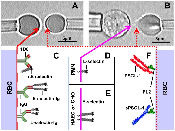
Figure 1. Micropipette adhesion frequency assay. A and B.
. Photomicrographs of a pair of RBCs (A) or a nucleated cell (left) and a RBC (right) (B) respectively held by two apposing pipettes. C-F. Composite of interacting molecules on respective cell surfaces. The left RBC in A was precoated with anti-E-selectin (1D6) that captured monomeric sE-selectin or precoated with goat anti-human Ig that captured dimeric E-selectin-Ig or L-selectin-Ig (C). The left nucleated cell in B was a PMN that expressed L-selectin (D) but could also be a HAEC or CHO cell that expressed E-selectin (E). Based on the data of this work, L-selectin on PMN is depicted as a monomer whereas E-selectin on HAEC and CHO cell is depicted as a dimer. The right RBCs in both A and B were precoated with anti-PSGL-1 mAb PL2 that captured dimeric membrane PSGL-1 or monomeric recombinant sPSGL-1 (F).

