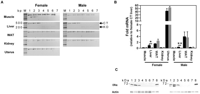
Figure 1. LERKO mice exhibit liver-specific downregulation of ERα.
A) Downregulation of the ERα transcript is confined to the liver. Real-time PCR screening of ERα transcript across various LERKO tissues revealed the ERα transcript was significantly downregulated in the liver, but not in the muscle, white adipose tissue (WAT), kidney and uterus (female). B) Hepatic ERα transcript levels were approximately 10 fold lower in LERKO (grey bars) as compared to CT (black bars). Data are represented as mean ±SEM of three individual mice. * = P≤0.05; ** = P≤0.01. C) Western blot analysis of CT and LERKO liver lysates confirmed a strong downregulation of the ERα protein (but not actin) in LERKO livers. Uterus samples of wild type and ERαKO mice served as positive and negative controls, respectively. Actin was used as loading control. Each lane represents a single animal sample. Lanes 1–3 = WT; 4–6 = LERKO; 7 = ERαKO uterus; 8 = CT uterus. It is notable that a second band is detected by the ERα antibody in the liver of male but not female mice. While it is difficult to identify the exact source of the second band, one possibility is that it represents male prominent ERα degradation products. In line with this, longer exposure reveals a double band also in one of the liver samples from female mice (data not shown).

