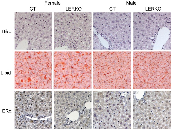
Figure 2. Immunohistochemical analysis of LERKO livers.
Male and female liver tissue sections were analysed for gross structural morphology, lipid content and ERα protein expression. Hematoxylin and eosin (H&E) staining indicated CT and KO animals had similar gross liver morphology. Lipid staining revealed that similar amounts of lipid droplets were present in CT and LERKO animals. Staining for ERα indicated a predominant hepatic nuclear localisation, which was decreased in LERKO mice of both sexes. Sections from three individual 6 month-old mice from each of the test groups were analysed. The figure shows representative sections.

