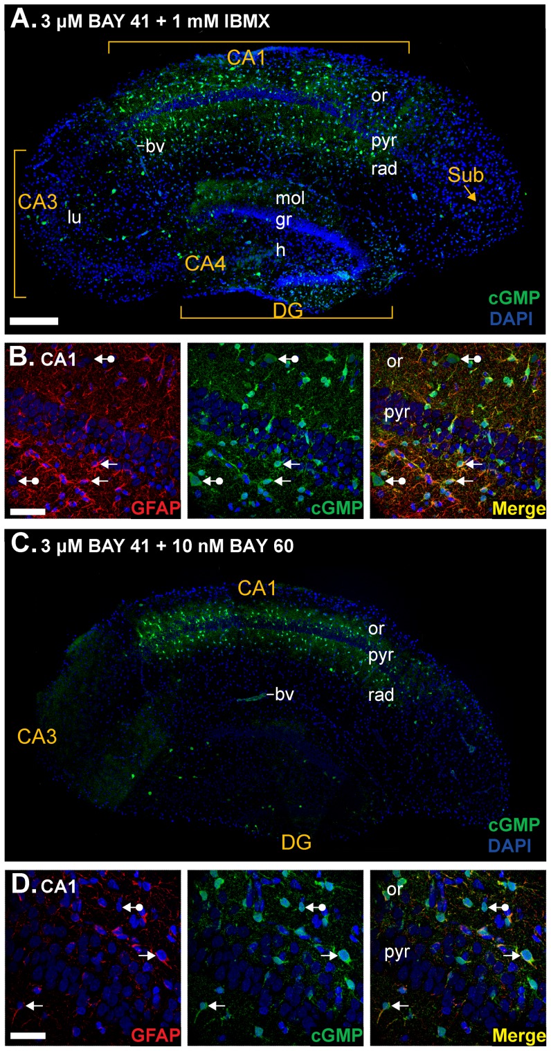
Figure 1. cGMP immunohistochemistry in immature rat hippocampal slices.
A,B. Sections from slices pre-treated with the non-specific PDE inhibitor IBMX (1 mM) and the allosteric enhancer of NO receptor-guanylyl cyclase BAY 41-2272 (3 µM) were immunolabelled for cGMP (green) and counterstained using DAPI (blue nuclei). A. Composite image of an entire hippocampal section. B. Double labelling for the astrocyte marker GFAP (red, left) and cGMP (green, middle) in the CA1 subfield. Colocalisation appears in yellow in the right-hand image. Arrows without tails indicate double-labelled cells and arrows with round tails, cGMP-positive, GFAP-negative cells. C, D. Slices were pre-treated with the PDE-2 inhibitor BAY 60-7550 (10 nM) instead of IBMX. Photographs are as in A and B. The number of GFAP-positive fibres in D appears to be fewer than in B, because of the oblique plane of section through the cell layer. Key: bv, blood vessel; DG, dentate gyrus; gr, granule cell layer; h, hilus; lu, stratum lucidum; mol, stratum moleculare; or, stratum oriens; pyr, stratum pyramidale; rad, stratum radiatum; Sub, subiculum. Scale bar in A = 200 µm (also applies to panel C); B = 30 µm; D = 50 µm.

