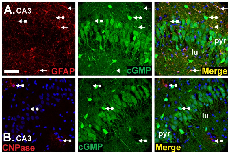
Figure 7. Double labelling for cGMP and glial cell markers.
Experimental conditions were as in Figure 4A, 5 and 6 (1 µM BAY 60-7550, 10 µM BAY 41-2272, 10 µM DEA/NO). Sections were doubled-labelled for cGMP (green) and a marker for astrocytes (GFAP; red in A) or oligodendrocytes (CNPase; red in B). Photographs show an area of the CA3 subfield and are representative of findings throughout the hippocampus. Arrows without tails: double-labelled cells and fibres; arrows with round tails, cGMP-positive, glial cell marker-negative cells; arrows with square tails, cGMP-negative, glial cell marker-positive cells. The key is as in Figure 1 legend. Scale bar in A = 50 µm and applies to both panels.

