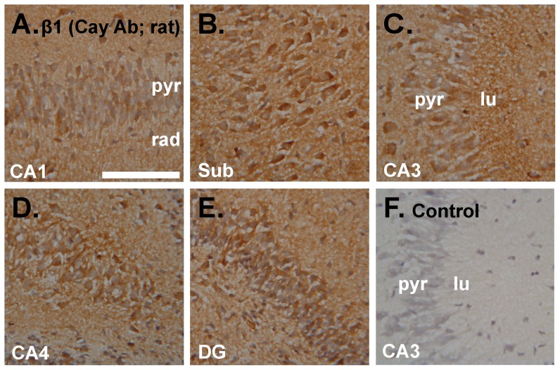
Figure 8. Immunoperoxidase staining using an antibody raised against the NO receptor-guanylyl cyclase β1 subunit.
A–E. Incubated immature rat hippocampal slices were sectioned and stained for the β1 subunit (brown) using a primary antibody from Cayman Chemical Company (Cay Ab). Photographs show an area of CA1 (A), the subiculum (sub; B), CA3 (C), CA4 (D) and the dentate gyrus (E). F. Control with the primary antibody omitted showing area CA3 (representative of the entire hippocampus). Tissues were fixed in 1% paraformaldehyde. See Figure 1 legend for key. Scale bar in F = 100 µm and applies to A–F.

