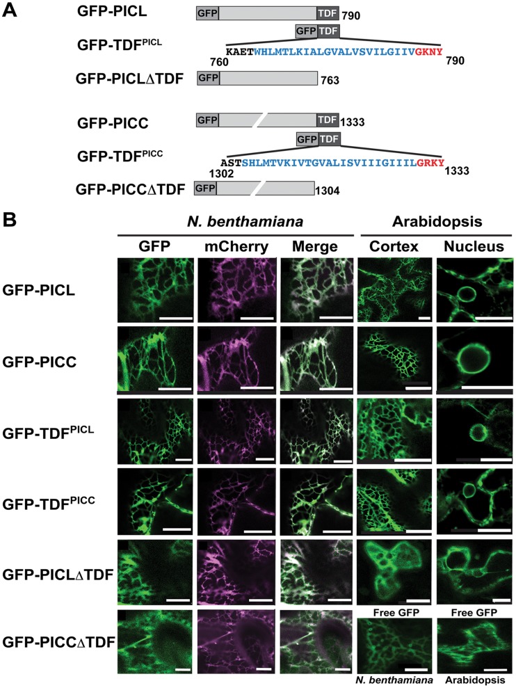
Figure 2. PICL and PICC are associated with the ER via their C-terminal transmembrane domain.
(A) N-terminally tagged GFP-fusion proteins used in this study. Amino acid sequence of the transmembrane domain and the C-terminal tail are shown in blue and red letters, respectively. Numbers indicate amino acid positions. Drawings are not to scale. (B) Confocal images showing localization of the fusion proteins indicated on the left in N. benthamiana and Arabidopsis leaf epidermal cells. Cytoplasmic localization of unfused GFP (“Free GFP”) in N. benthamiana and Arabidopsis are shown as controls (bottom right). Scale = 10 µm.

