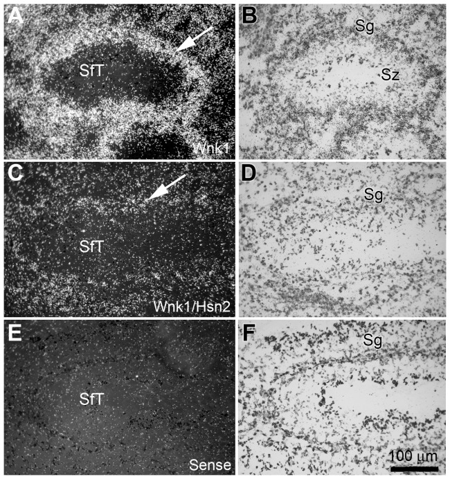
Figure 12. Wnk1 and Wnk1/Hsn2 expression in the testis of adult mice.
(A, C and E) Tissue sections prepared from mouse testis were used in ISH detections with anti-sense Wnk1, anti-sense Wnk1/Hsn2 and sense Wnk1/Hsn2 (negative control) riboprobes, respectively. (B, D and F) Corresponding sections were stained with cresyl violet. Arrows in A and C indicate the increased expression of Wnk1 in the wall of the seminiferous tubule spermatogonia cell layers, in comparision to Wnk1/Hsn2. Sc = spermatocyte; SfT = seminiferous tubule; Sg = spermatogonia; Sz = spermatozoa.

