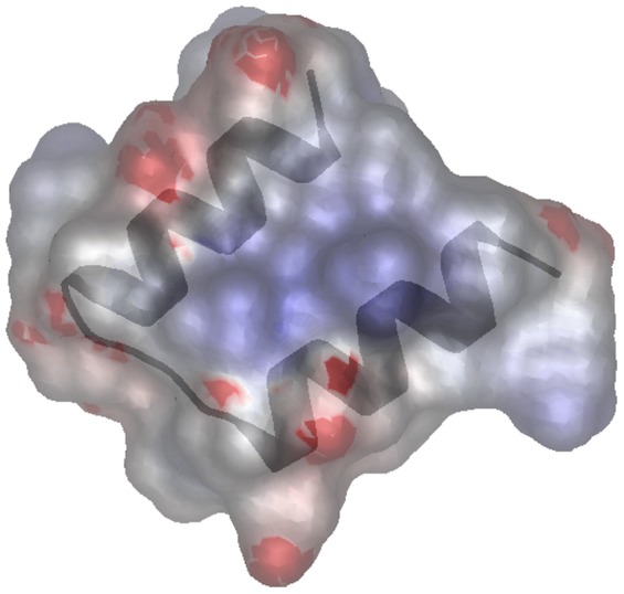
Figure 2. Three-dimensional structure prediction of the SHOCT domain of UniProtKB B0PET9.1 (residues 20–50) generated with QUARK using the default parameters[12] and viewed using MarkUs [44].

Surface electrostatic potential for the model is calculated using the program GRASP2 [45] accessed through MarkUs. The positively charged areas of the protein surface are shown in blue, and negatively charged areas in red, the two alpha helices are overlaid in grey.
