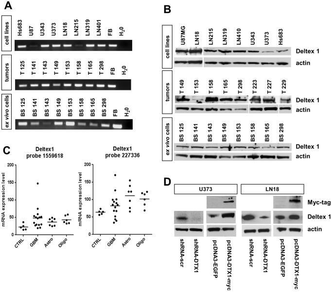
Figure 1. DTX1 is expressed in glioblastomas, ex vivo cells and established glioma derived cell lines.
(A) Semi-quantitative RT-PCR probing for exon 1 of DTX1 in established glioma derived cell lines, glioma tumor biopsies and ex vivo cell lines. Ex vivo cell lines were derived from tumor biopsies as indicated by the numbering and were maintained as low passage cultures. Fetal brain (FB) was used as positive control. (B) Western Blot analysis of glioma derived cell lines, glioma tumor biopsies and ex vivo cell lines probing for DTX1 and β-actin. (C) Microarray gene expression analysis of tumors and control tissue. Two non-diseased normal brain samples and three normal human astrocyte cultures were used as controls (ctrl), 15 GBMs, seven astrocytomas and six oligodendrogliomas were analyzed. Dots represent individual specimens, average expression values are shown as lines. Results for two independent probes on the chip detecting DTX1 mRNA are shown. (D) Western blot analysis of transfected cell lines U373 and LN18 probing for DTX1, Myc-tag and β-actin demonstrating DTX1 over-expression and down regulation as indicated according to the genotype.

