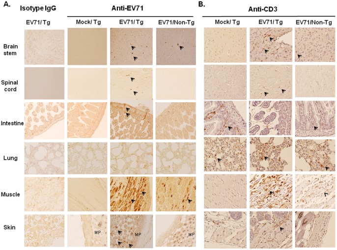
Figure 4. In situ EV71 distribution in hSCARB2-Tg mice.
Seven-day-old hSACRB2-Tg and non-Tg mice infected with 3×10⁴ pfu of EV71 5746 s.c. were sacrificed on Day 7 post-infection. Uninfected hSCARB2-Tg mice were used as the negative control. Waxed sections of the brainstem, spinal cord, intestine, lung, biceps femoris muscle, and lower back skin were prepared and IHC stained with (A) Mab979 antibody or isotype mouse IgG and (B) the anti-CD3 antibody. All pictures were taken at 200X magnification. Viral particles or T lymphocytes in the sections are indicated with arrows. The melanin pigments (MP) pale-stained by Mab979 antibody in the section of skin tissue was observed.

