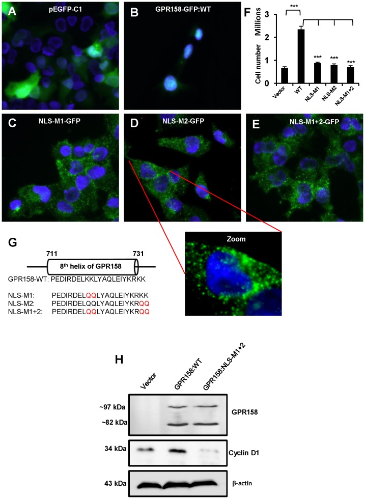
Figure 7. Role of the bipartite NLS in nuclear localization and cell proliferation.
(A, B, C, D and E) The fluorescent images were captured of cells transfected with either vector (A) or GPR158-GFP plasmid (B) or NLS-M1-GFP plasmid (C) or NLS-M2-GFP plasmid (D) or NLS-M1+2-GFP plasmid (E). The representative images from two independent transfection experiments are shown. 3 days post-transfection, the cells were fixed with 4% PFA, washed with PBS and the slides were mounted using VECTASHIELD with DAPI and viewed using Nikon Eclipse Ti-E fluorescence microscope. The merged images show GPR158-GFP as green and nuclear stain DAPI as blue. The zoom panel indicates the magnified image of an indicated cell. (F) After 3 days of transfection, the cells transfected with above indicated plasmids were trypsinized and counted using trypan blue dye in a hemocytometer chamber. The data represent mean ± SEM of two independent experiments. (G) Schematics showing amino acid sequence corresponding to the bipartite NLS, a part of 8th helix of GPR158. The mutated amino acids are shown in color red in indicated NLS mutant constructs. (H) TBM-1 cells were transfected with either GPR158 wild type or NLS-M1+2 or GFP vector alone plasmid using Lipofectamine LTX reagent. After 3 days of transfection, the cell lysates were prepared using RIPA buffer and the western blotting for the detection of GPR158 and cyclin D1 was performed using appropriate antibodies. The same membrane was striped and reprobed for β-actin for loading control. The data represent two independent experiments performed in triplicate.

