
Figure 5. Staining for VCAM1 in the liver.
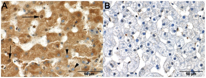
Marked granular cytoplasmic immunoreactivity with the presence (arrows) and absence (arrowheads) of immunoreactivity in the nuclei of hepatocytes in a liver sample taken from a dog with an intrahepatic portosystemic shunt (Figure 5 A). The cytoplasm of hepatocytes in a liver sample from a dog with an extrahepatic portosystemic shunt (EHPPS) show no immunoreactivity. Nuclei in this liver occasionally demonstrate weak immunoreactivity (Figure 5 B).