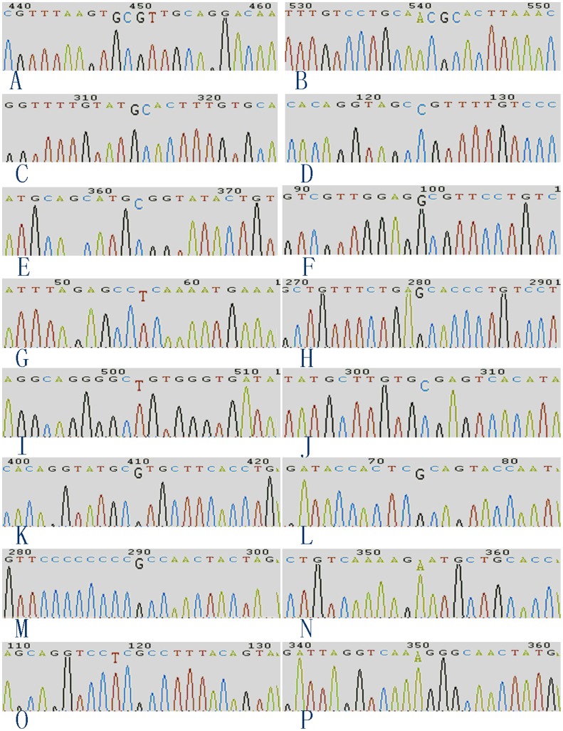
Figure 1. Chromatograms of sequence variations.
The sequence variations were enlarged to facilitate observation. In B, D, E, F, I and J, the reverse complement of the sequence variations are displayed because the sequences were obtained using downstream primers. G, guanine; C, cytosine; A, adenine; and T, thymine.

