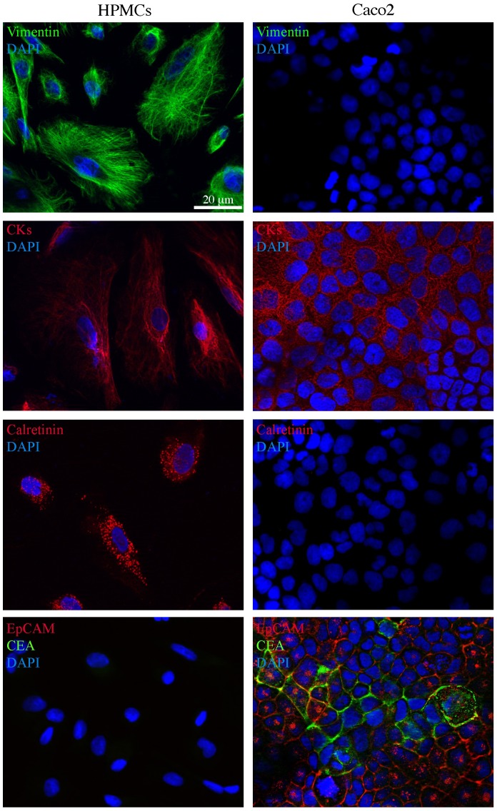
Figure 3. Immunofluorescence characterization of human peritoneal mesothelial cells from peritoneal washes of gastric and colon cancer patients.
Primary cultures of human peritoneal mesothelial cells (HPMCs) were isolated from peritoneal washes as described in materials and methods. Caco2 colon cancer cells were used as a control. Immunofluorescence analysis using antibodies directed against mesothelial (vimentin, CK8 and CK19 cytokeratins and calretinin) and epithelial (EpCAM and CEA) markers shows that HPMCs are positive for vimentin and cytokeratin staining, that appears as perinuclear bundles of filaments, as well as for the hot-spotted calretinin signal, but are negative for the plasma membrane EpCAM staining and for the intracellular and surface CEA signal. Caco2 cells are positive for cytokeratins and double positive for the EpCAM and CEA epithelial markers visible on the cell surfaces (EpCAM, green signal) or on the plasma membranes and in intracellular spots (CEA, red signal). Nuclei were stained with DAPI. Bar: 20 µm.

