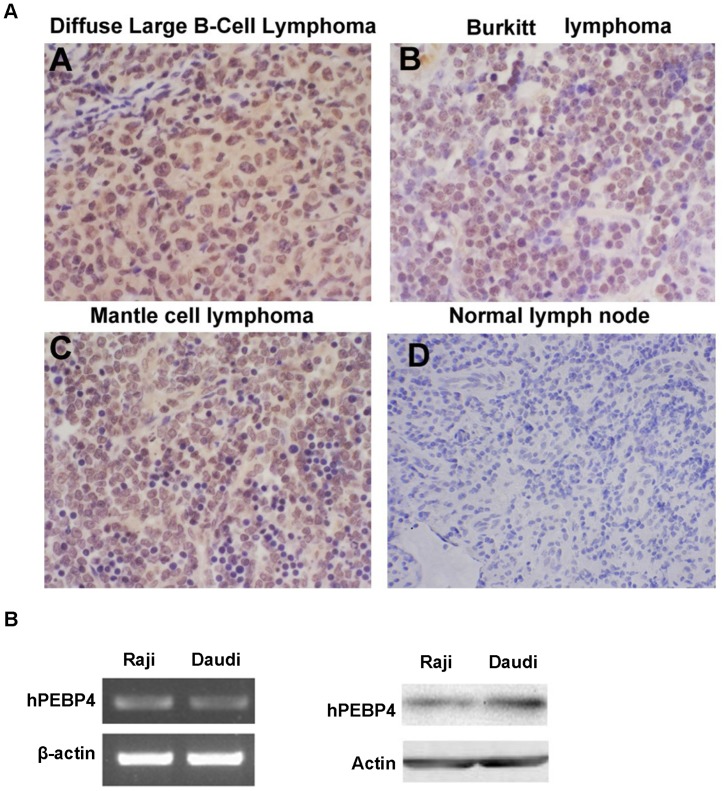
Figure 1. hPEBP4 is highly expressed in human lymphoma.
A. Representative results of immunohistochemical staining of hPEBP4 protein (Yellow) in one sample with no signal in the normal lymph node (panel d) but positive staining in lymphoma samples (panels a–c). Photos were taken under×200 magnifications. B. RT-PCR (left) and Western blot analysis (right) of hPEBP4 expression in B-NHL cell line.

