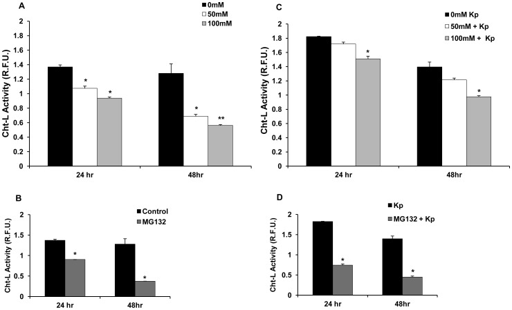
Figure 3. Ethanol suppresses 20S Cht-L proteasome activity in murine primary peritoneal macrophages. A.
Murine peritoneal macrophages (2.5×10⁵ cells/ml) were treated with 50 mM or 100 mM EtOH for 24 h or 48 h. 0 mM (control) cells received no treatment. Cell lysates obtained at the indicated time points were analyzed for 20S Cht-L proteasome activity by fluorigenic assay as described in Methods. B. Peritoneal macrophages were treated with MG132 (5 uM), as a positive control of proteasome suppression. Cell lysates were obtained at the indicated time points and analyzed as described in Panel A. C. Ethanol-treated peritoneal macrophages were stimulated with heat-killed Kp, 6 hours after initial EtOH exposure. Cell lysates obtained at indicated time points were analyzed for proteasome activity as described in panel A. D. MG132-treated peritoneal macrophages were stimulated with Kp as described in Panel C. Cell lysates were obtained at the indicated time points and analyzed for proteasome activity as in Panel A. Proteasome activity data plotted as mean relative fluorescence units (R.F.U.) ± SEM (three replicates per experiment) and is representative of three independent experiments. Higher R.F.U. values indicate higher proteasome activity. *p<0.05 vs. control at the respective time point. **p<0.05 vs. control and 50 mM EtOH at the respective time points.

