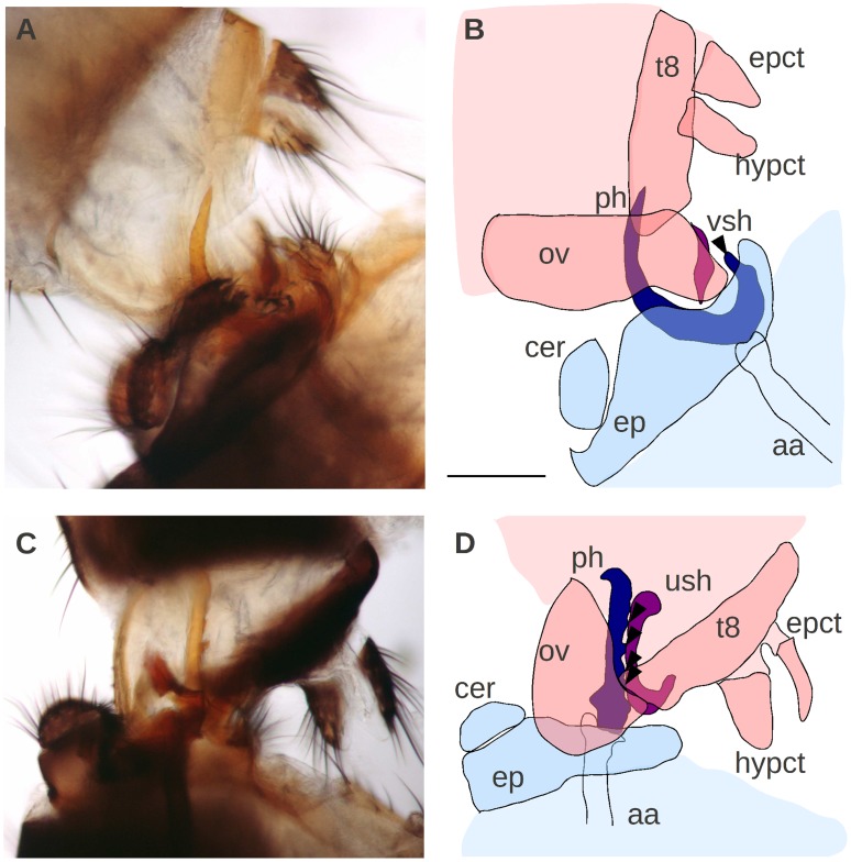
Figure 5. Copulation anatomy of D. orena (A–B) and D. erecta (C–D).
Male and female organs are depicted in blue and pink, respectively, with contacting species-specific structures in dark colors. Note that phallic hooks and spines (arrowheads) contact female vaginal shields during copulation; aa: aedeagal apodeme; cer: cercus; ep: epandrium; epct: epiproct; hypct: hypoproct; ov: oviscapt; ph: phallus; t8: tergite 8; ush: uterine shield; vsh: vulval shield. Note that the male surstyli that grasp the female oviscapt at the ventro-distal margin and the phallic paraphyses were not reproduced in the schematic drawings (B, D) for the sake of clarity. Scale bar is 1 mm.

