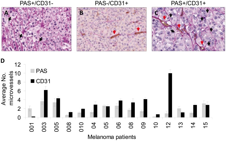
Figure 1. Characterization of vessel with PAS-CD31 dual staining (endothelial or vessel like channels – VM) in paraffin sections of melanoma patients.
(A) PAS-positive VM channels with no endothelial marker CD31 staining, black arrows indicate VM channels; (B) PAS-negative patterns and CD31-positive staining; red arrows indicate endothelial channels; (C) PAS-positive and CD31-positive channels; (D) a summary of microscopic vessels observed. Microscopic evaluation was done blindly by two pathologists. The data represents average values as seen in 10 high power fields per sample.

