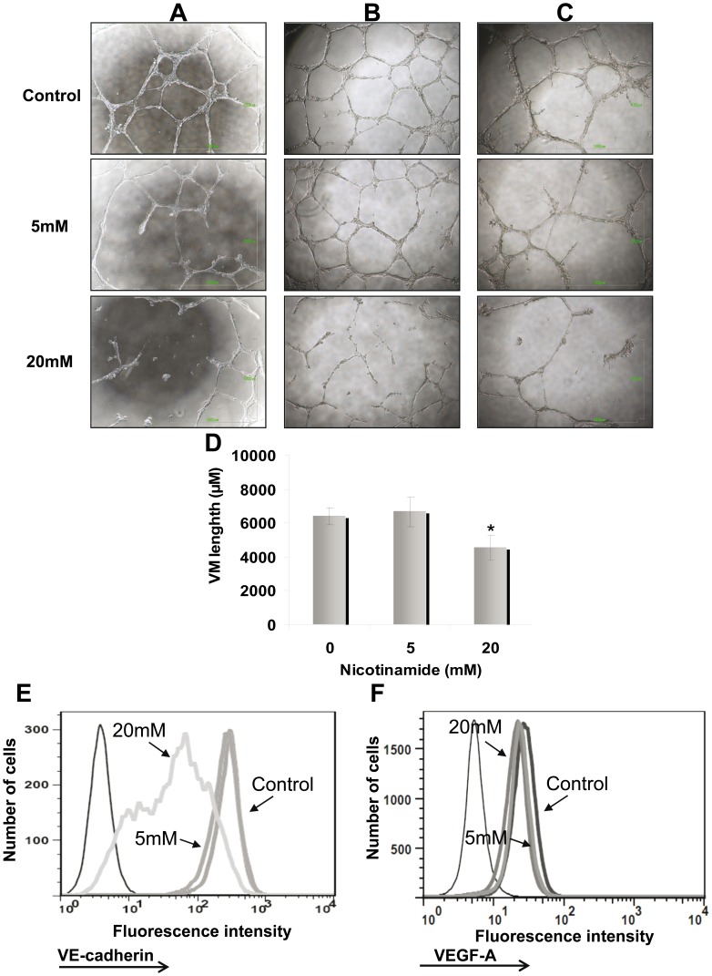
Figure 4. Nicotinamide (NA) abrogates vasculogenic mimicry.
(A) NA was added 24 h after capillary-like structures were developed (destruction of VM formation). NA was added in concentrations of 5 mM and 20 mM as indicated for additional 24 h. Afterwards, microphotographs were captured; (B) HAG was cultured in the presence of NA (same concentrations as A) for 1 month (prevention of VM formation). Subsequently, the treated cells were cultured on Matrigel to enable VM formation. The images were captured after 24 h; (C). Prolonged inhibition of VM formation by NA treatment. The cells were incubated with NA for 72 h, re-plated without NA for 1 month, followed by VM testing. Microphotographs were capture 24 h after plating. Each picture (A–C) was derived from one representative experiment out of three performed. Each experiment was performed in duplicates; (D) Tube formation was quantified using the ImageJ analyze skeleton PlugIn. Figure showed the average length calculated for each sample out of all image captured in all three experiments performed. Statistical significant was tested with 2-tailed paired t-test. (E) Effect of NA on VE-cadherin expression in the HAG cells cultured for 1 month in the presence of NA. (F) Effect of NA on VEGF-A expression in the HAG cells cultured for 1 month in the presence of NA. Black thin line histogram represents cells stained with isotype control; other histograms represent cells treated either with vehicle or various NA concentrations, as indicated in the figure. Figure shows representative experiments out of three performed.

