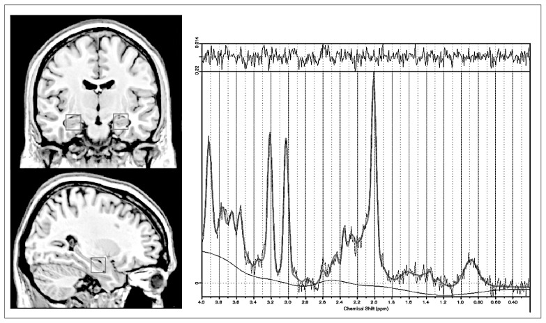
Fig. 1.
Magnetic resonance images showing (left) the placement of the volumes of interest in a coronal and sagittal section of the medial temporal region and (right) a representative 1HMRS postprocessed with LCModel: PRESS acquisition at 3 T, with a repetition time of 2000 ms and echo time of 38 ms.

