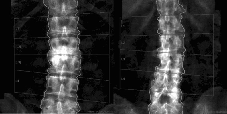
Fig. 2.
Examples of spinal degenerative changes. Examples of what were considered as “obvious” degenerative changes on the DXA images are presented here. The image on the left is a 70-year-old man in whom focal abnormalities at L2 and L3 are apparent. The individual vertebral body T-scores are consistent; from L1 to L4 these values are +1.1, +4.5, +4.2 and +1.7; thus, a >1 T-score difference is present between adjacent vertebrae. The 86-year-old female on the right has apparent degenerative changes at L2, L3 and L4 with obvious osteophytes at L2 and L3. T-score differences between adjacent vertebrae were observed with values from L1 through L4 being +0.2, +1.7, +3.5 and +4.1, respectively

