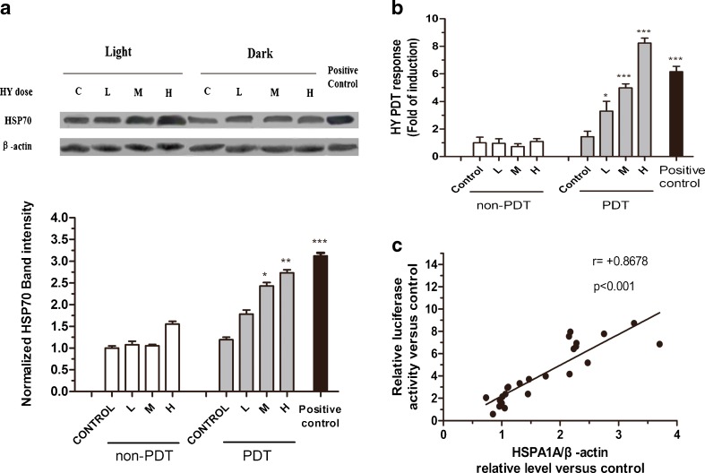
Fig. 5.
MCF-7 luciferase cells respond to PDT mediated by various concentration Hyp with increased levels of HSPA1A and relative luciferase activity. H high dose, M middle dose, L low dose. The MCF-7 luciferase cells were treated by PDT mediated by various concentration Hyp (H, 0.125 μM; M, 0.0625 μM; L, 0.03125 μM) with a light dose of 3.87 J/cm2, and then recovered at 37 °C for the indicated times. The untreated cells were used as control. Heat shock treatment was performed as positive control. a HSPA1A levels detected by Western analysis after PDT treatment. β-actin was used as the loading control. Levels of HSPA1A and β-actin were determined by densitometric analysis. b Relative luciferase activity of MCF-7 luciferase cells treated by PDT mediated by various concentration Hyp was measured using the luciferase assay system. c Correlation between relative luciferase activity and relative level of HSPA1A was evaluated using the Spearman rank test. Data represent mean ± SD of three individual experiments

