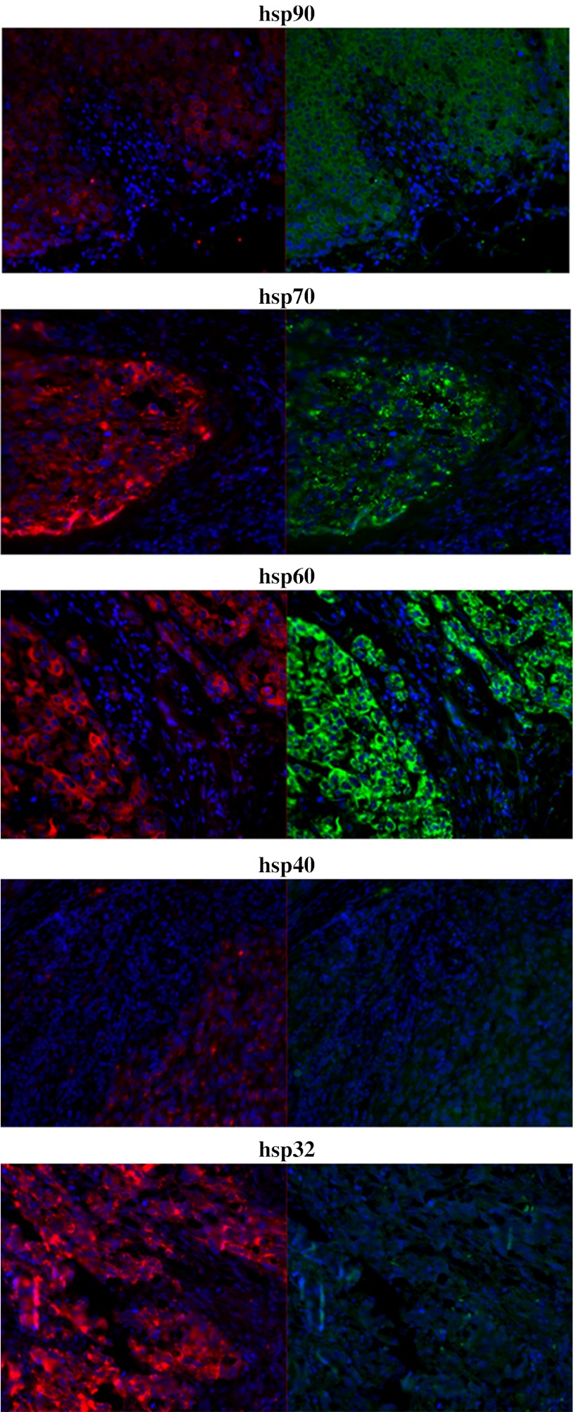
Fig. 5.
Fluorescence microscopy of heat shock protein expression in melanoma tumour tissue. Hsps 90, 70, 60, 40 and 32 were increased in expression in MelanA+ cells relative to MelanA− cells. Formalin-fixed paraffin-embedded melanoma tissue sections were stained for hsp expression with fluorescent antibodies and analysed using fluorescence microscopy. Representative images shown. Red MelanA (marker for melanoma cells), green hsp (marker for hsp), blue DAPI DNA stain (marker for cell nucleus), 20× magnification

