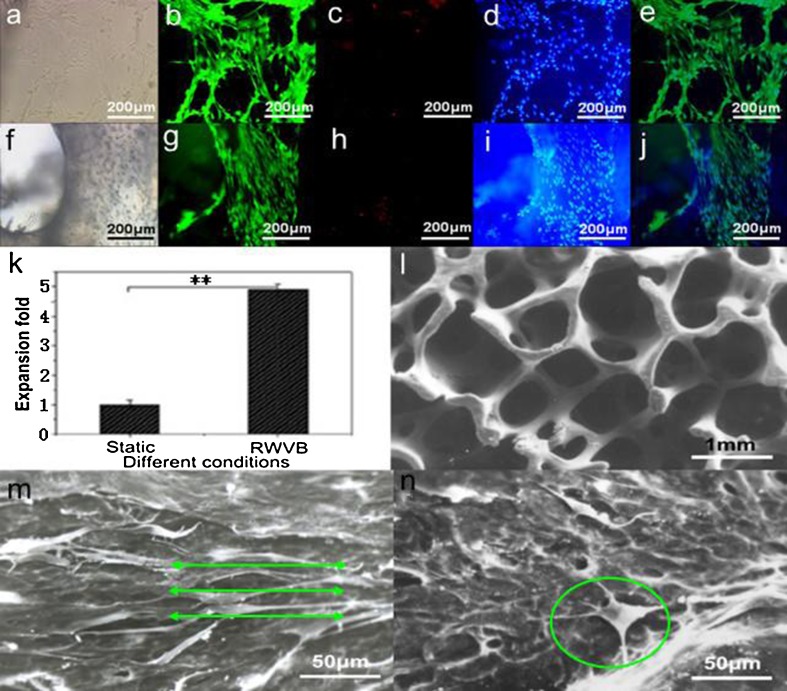
Fig. 7.
Live/dead assay of passaged osteoblasts at day 7 and cell viability, expansion, and morphology of osteoblast/bio-derived bone scaffold constructs after 7 days of fabrication. a Bright field, b live cells stained with calcein AM, c dead cells stained with PI, d blue nuclei of live cells stained with Hoechst 33342, e overlayed by b–d; f bright field, g live cells stained with calcein AM, h dead cells stained with PI, i blue nuclei of live cells stained with Hoechst 33342, j overlayed by g–i; a–j ×100. k Comparison of cell expansion for osteoblasts in different culture fashions. l SEM micrographs of vacant scaffold. SEM micrographs of scaffolds cultured with osteoblasts in RWVB (m, green lines showed the cellular direction for osteoblast spreading affected by hydrodynamic stimulation) and static T-flask (n, osteoblast marked with green circle did not show typical phenotype) after 1 week of fabrication

