Fig. 8.
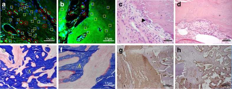
Laser confocal micrograph, HE stained, Masson stained and type I collagen assay of histological sections of rabbit radial defects repaired with tissue-engineered bone constructs after 8 weeks. Laser confocal microscope picture in the RWVB repair group (a) and the static repair group (b): sun with rays ectogenous osteoblasts, square endogenous osteoblasts, 600×; HE stains in the RWVB repair group (c) and the static repair group (d), right-pointing pointer osteoclasts, 100×; Masson stains in the RWVB repair group (e) and the static repair group (f), triangle new bones, 600×; type I collagen assays in the RWVB repair group (g) and the static repair group (h), 100×

