
Figure 2.
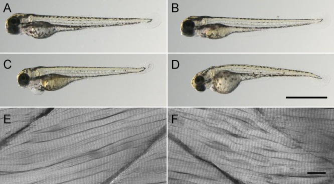
Morphology of control and morphant larvae. Microscopy photographs of 4-dpf wild-type (A) and control-injected (B) larvae. (C and D) Splice- and translation-blocking MO-injected morphants, respectively. Both types of morphants had signs of cardiac edema. (E and F) Confocal microscopy of a control (E) and a splicing morphant (F) stained with rhodamine phalloidin. Bars: (A–D) 1 mm; (E and F) 10 µm.