Figure 3.

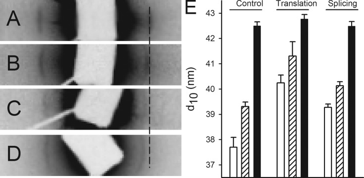
X-ray diffraction of control and morphant larvae. The equatorial region of small angle x-ray diffraction patterns recorded from control-injected (A) and splice- and translation-blocking morphants (B and C) at 4 dpf. The muscles were mounted at optimal length (Lopt). The equator in the pattern is oriented horizontally, and the position of the 1.0 reflection (corresponding to 37.9 nm) in the control is indicated with a broken line. (D) A recording from a living, anesthetized control larva in E3 medium. (E) The spacing of 1.0 reflection in the different groups at Lopt (open bars, sarcomere length 2.1–2.2 µm), slack (Ls, hatched bars, ∼1.7 µm), and in a living state (shaded bars). Statistical analysis (n = 3–6 in each group, P < 0.05; one-way ANOVA, and Holm-Sidak method for multiple comparisons) revealed that shortening (Lopt vs. Ls) gives a significant increase (P < 0.05) in spacing of controls and translation-blocking morphants. Living larvae had a wider spacing in all groups compared with mounted larvae (at Ls or Lopt). Morphants (translation and splicing) had a wider spacing than controls at Lopt and for translation also at Ls. No differences were detected between the living larval groups. Error bars indicate mean ± SEM.
