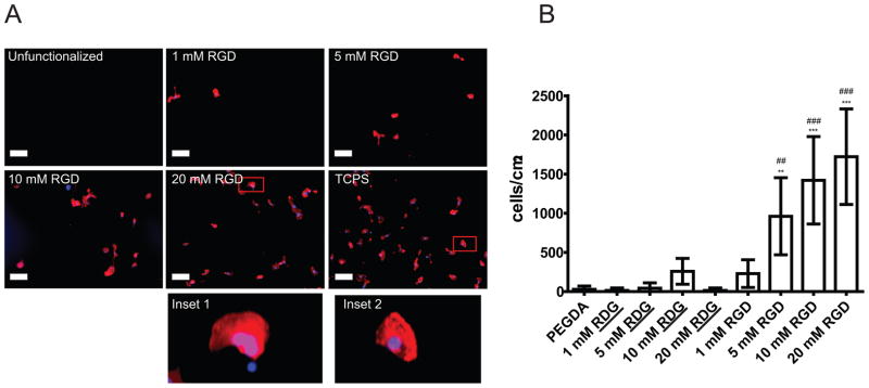
Figure 3. Increasing the surface density of RGD promotes cell attachment.

A) Representative images demonstrating increasing levels of RGD peptide to promote cell attachment on PEGDA 3400/20% hydrogels. Cells were plated in EP media for 24 hours, followed by fixation, staining and analysis of cell attachment. Red: TRITC-Phalloidin. Blue: DAPI. Negative control: Unfunctionalized PEGDA. Positive control: Tissue Culture Polystyrene (TCPS). Scale bar: 100 μm. Inset 1 (20 mM RGD surface) and inset 2 (TCPS) demonstrate similar cell spreading.
B) Cell attachment is RGD concentration dependent. In EP media, non-functionalized hydrogels made with PEGDA 3400/20% and hydrogels functionalized with scrambled peptide RDG presented no attachment of HCECs. Hydrogels functionalized with RGD had increased number of HCECs attached after 24-hours culture when the concentration of RGD increased. Error bars: SEM (*: vs flat. #: vs scrambled. *** P ≤ 0.001; ** 0.001 < P ≤ 0.01; * 0.01 < P ≤ 0.05).
