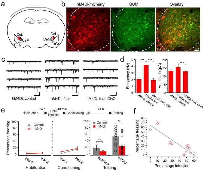
Figure 4. Synaptic potentiation onto SOM+ neurons in CeL is required for the formation of fear memory.
The SOM-IRES-Cre mice were used in these experiments. (a) A schematic experimental design. The hM4Di-mCherry is expressed in SOM+ neurons in CeL (shown in red) by viral infection. (b) An example image of the SOM+ neurons in CeL infected with the AAV-DIO-hM4Di-mCherry virus. Left: expression of hM4Di (red) was detected based on the intrinsic fluorescence of mCherry. Middle: SOM+ neurons (green) were recognized by an antibody against SOM. Right: Only SOM+ neurons expressed hM4Di, as indicated by the overlapping of red and green signals. Dashed lines mark the border of CeL. Coronal sections. (c) Representative mEPSC traces recorded from SOM+ CeL neurons expressing hM4Di-mCherry, in the following groups: “hM4Di, control” (control), “hM4Di, fear” (fear conditioning), and “hM4Di, fear, CNO” (fear conditioning with CNO pre-treatment). For the fear conditioning groups, acute brain slices were prepared 3 hr following fear conditioning. Calibrations: 20 pA and 500 ms. (d) Left: CNO pre-treatment suppressed the fear conditioning-induced increase in mEPSC frequency in SOM+ neurons that expressed hM4Di (control: 2.02 ± 0.35 Hz, n = 11 cells, 3 animals; fear: 4.18 ± 0.33 Hz, n = 15 cells, 3 animals; fear, CNO: 1.93 ± 0.22 Hz, n = 12 cells, 3 animals; ***P < 0.001, bootstrap with Bonferroni correction). Right: CNO pre-treatment also suppressed the fear conditioning-induced increase in mEPSC amplitude in SOM+ neurons that expressed hM4Di (control: 13.1 ± 0.81 Hz, n = 11 cells, 3 animals; fear: 15.24 ± 0.51 Hz, n = 15 cells, 3 animals; fear, CNO: 12.75 ± 0.47 Hz, n = 12 cells, 3 animals; ***P < 0.001, bootstrap with Bonferroni correction). (e) Top: A schematic experimental procedure. Bottom left & middle: freezing behavior during habituation and conditioning. Control: SOM-IRES-Cre mice that received AAV-DIO-GFP injection bilaterally into CeL. hM4Di: SOM-IRES-Cre mice that received AAV-DIO-hM4Di injection bilaterally into CeL. Freezing responses were similar for the two groups at the end of conditioning (control, 14.2 ± 4.9%, n = 9 animals; hM4Di, 16.6 ± 6.1%, n = 15 animals; P > 0.05, t-test). Bottom right: the hM4Di mice showed impaired fear memory recall compared with control (control, 52.36 ± 8.76%, n = 9 animals; hM4Di, 23.24 ± 6.05%, n = 15 animals; t = 2.82, DF = 22, **P < 0.01, t-test). The two groups did not differ significantly in their baseline freezing levels (control, 17.56 ± 9.62%, n = 9 animals; hM4Di, 6.67 ± 2.37%, n = 15 animals; P > 0.05, t-test). (f) The freezing level of individual animals correlated with the extent of infection, measured as the fraction of SOM+ CeL neurons that expressed hMD4i-mCherry (R2 = 0.76, F(1,13) = 45.62, grey line; P < 0.001 by a linear regression; n = 15 animals). The extent of infection for the left and right CeL was averaged. Error bars, s.e.m.

