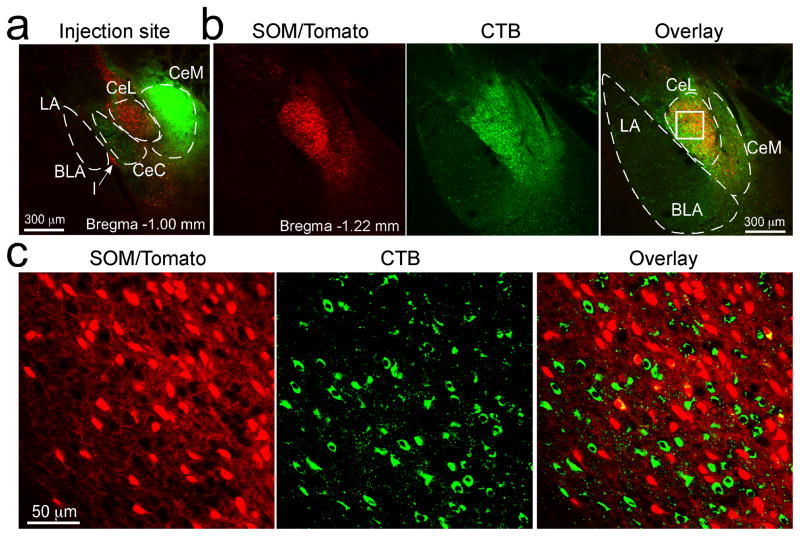
Figure 5. SOM+ CeL neurons do not project to CeM.
(a) CTB (green) was injected into the CeM of a SOM-IRES-Cre/Ai14 mouse (similar results were obtained from 2 animals). Coronal brain section (at ~Bregma −1.00 mm). (b) Images of a brain section from the same mouse (at ~Bregma −1.22 mm). Left, the SOM+ neurons expressed tdTomato (red). Middle, extensive labeling by CTB (green) was seen in CeL. Right, overlay; the box in CeL marks the region shown in c at higher magnification. (c) Higher magnification images of the boxed region in CeL in b. The vast majority of the CTB-labeled neurons (green) in CeL were not SOM+ (red; see overlay in right).

